Datasheet is currently unavailable. Try again or CONTACT US
Datasheet Protocol

WM1341D Viable Cells

7 References
WM1341D-01-0001 WM1341D-01-0005 WM1341D-01-0010
1 million cells 5 x 1 million cells 10 x 1 million cells
Frozen Cell Suspension Frozen Cell Suspension Frozen Cell Suspension
WB, IHC, IF, Cellular Assay, Functional Assay, Multiplex
Human
$1,089.00 /Per Item
$3,629.00 /Per Item
$5,445.00 /Per Item
1 million cells $1,089.00
5 x 1 million cells $3,629.00
10 x 1 million cells $5,445.00
In stock
Ships next business day
Out of stock
Call for availability
Out of stock
Call for availability
Shipping info:

$50.00 to US & $70.00 to Canada for most products. Final costs are calculated at checkout.

Product Details

WM1341D Viable Cells - WM1341D-01-0001
Melanoma, patient derived tumor, tumor models, skin cancer, xenograft
Human

Target Details

Cells are sterile, validated by short tandem repeat profiling, and are tested as negative for mycoplasma. It is recommended that cell lines are tested for mycoplasma contamination and short tandem repeat (STR) profiling every 10 passages or each time a frozen seed stock is made. See cell culture protocol for additional details.

Application Details

Cellular Assay, Functional Assay, IF, IHC, WB, Multiplex  - View References
The key applications of these cell lines include genetic studies, xenograft production, drug testing, and drug target discovery. These cell line models can be used in various biological assays, and for identifying critical target genes, and cell signaling pathways.

Cell Line Data

Human Melanoma
Viable Cells
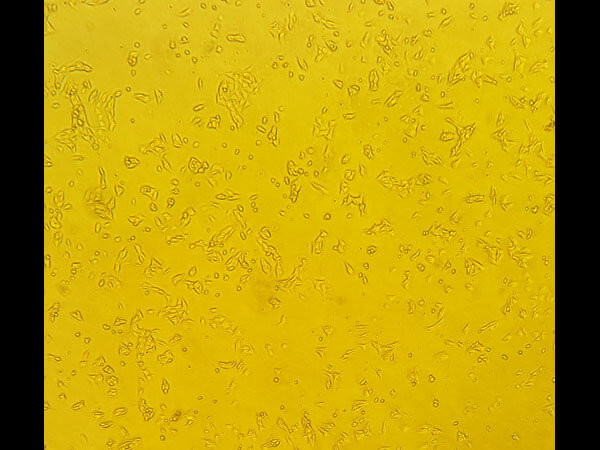
epithelial square and triangle
V600R
WT
WT
WT
Tumor Specialized Media with 2% HI-FBS
Cells should be maintained between 30 – 95% confluence in tumor specialized medium with 2% FBS; split cultures 1:3 every 10 days using 0.25% trypsin/EDTA.
36°C with 5% CO2

Formulation

1.0 million cells/mL Count By Hemocytometer
None
None
None

Shipping & Handling

Dry Ice
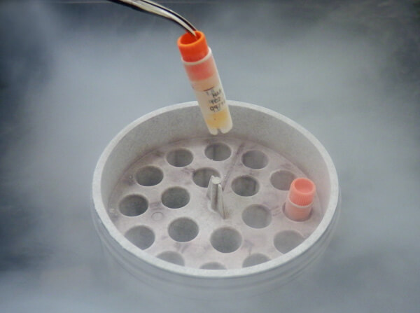
Cells are frozen with 90% FBS/10% DMSO solution at about 1x10^6 cells/ml. Store vial in liquid nitrogen upon arrival.
Expiration date is two (2) years from date of receipt.
WM1341D is a tumorigenic (VGP) primary melanoma cell line with competence for metastasis. These cells display epithelial square and triangular morphology in culture. This cell line features the specific V600R (Val600Arg) mutation at codon 600 in the BRAF gene. The V600R mutation results in an amino acid substitution at position 600 in BRAF, from a valine (V) to an arginine (R). This mutation occurs within the activation segment of the kinase domain. Mutations at V600 result in increased kinase activity and are transforming in vitro. WM1341D cells produce xenograft tumors when injected into immunocompromised mice.
(). Alpha-Enolase (ENO1) Correlates with Invasiveness of Cutaneous Melanoma—An In Vitro and a Clinical Study. Diagnostics (Basel).
Applications
IF, Confocal Microscopy; Other
(). Changes in Biomechanical Properties of A375 Cells Due to the Silencing of TMSB4X Expression Are Not Directly Correlated with Alterations in Their Stemness Features. Cells.
Applications
IF, Confocal Microscopy; Other
(). PARP1 as a Marker of an Aggressive Clinical Phenotype in Cutaneous Melanoma—A Clinical and an In Vitro Study. Cells.
Applications
IF, Confocal Microscopy; qRT-PCR, RT-PCR; WB, IB, PCA
(). The origin of the expressed retrotransposed gene ACTBL2 and its influence on human melanoma cells' motility and focal adhesion formation. Sci Rep.
Applications
IHC, ICC, Histology; WB, IB, PCA
(). Knockout of ACTB and ACTG1 with CRISPR/Cas9(D10A) Technique Shows that Non-Muscle β and γ Actin Are Not Equal in Relation to Human Melanoma Cells' Motility and Focal Adhesion Formation. Int J Mol Sci.
Applications
IF, Confocal Microscopy
(). Thymosin β4 regulates focal adhesion formation in human melanoma cells and affects their migration and invasion. Front Cell Dev Biol.
Applications
IF, Confocal Microscopy; Other; WB, IB, PCA
(). Varying effects of EGF, HGF and TGFβ on formation of invadopodia and invasiveness of melanoma cell lines of different origin. Eur J Histochem.
Applications
IF, Confocal Microscopy; WB, IB, PCA; Multiplex Assay

How can I ensure that the cells will be viable after resuspension?

Complete adherence to the protocol is necessary to ensure the viability of the cells. Cells should be maintained between 30-95% confluence in Tumor Specialized media with 2% FBS (heat inactivated) unless otherwise noted in cell line specific instructions.

How should I handle the cells before culturing?

Before thawing, frozen cells can be stored in liquid nitrogen or at -80°C only. To thaw the melanoma cells, move the vial from liquid nitrogen to dry ice immediately. Melanoma cells should be thawed quickly. Freeze thaw cycles can damage / kill the cells and should be avoided. Centrifuge the cells at 1500 rpm (500 x g) for 5 minutes at room temperature. Discard the supernatant. Resuspend cell pellet in 5 mL of Tumor Specialized Media with FBS (heat inactivated) and transfer to a T-25 flask. Place the flask in a humidified incubator (5% CO2) at 36°C overnight. Check the flask after 24 hours for attachment of cells to the flask. If cells are attached, remove the media and add fresh media to the flask.

Which media should I use to grow melanoma cells?

Tumor specialized Media with 2% FBS is preferred media for melanoma cell lines. However, alternative media (not preferred) recommendations include: 1. DMEM with 5% FBS; 2. RPMI with 5% FBS.

Do you have recommended sources for Tumor Specialized Media components?

The recommended manufacturer for each component of the Tumor Specialized Media are: MCDB-153 (Sigma-Aldrich, item# M-7403), Leibovitz’s L-15 (Sigma-Aldrich, item# L1518), FBS (heat inactivated) (Rockland, item# FBS-010-0100), Calcium Chloride (Sigma-Aldrich, item# C5670), Sodium Bicarbonate [Added to MCDB-153 preparation] (Sigma-Aldrich, item# S5761). Please consult the protocol for detailed instructions on the preparation of the media.

What is the optimal growth temperature for melanoma cells?

Melanoma cells grow best at 36°C (not 37°C) with 5% CO2. These incubation conditions must be followed exactly to ensure the viability and growth of the cells.

What is the recommended media for freezing viable melanoma cells?

Freezing media containing 90% FBS and 10% DMSO. Thawed cells should not be frozen again. Melanoma cells should be frozen slowly but thawed quickly.

What is the source of the melanoma cell lines?

The melanoma cell lines were established from patient-derived tumors. Information about the patients or treatments received is not available.

Are melanoma cell lines immortalized?

Melanoma cell lines are considered immortalized because they can replicate indefinitely.

Invalid lot number

If you need help finding your CoA, contact us

No test method can provide total assurance that the hepatitis B virus, hepatitis C virus, human immunodeficiency virus, or any other infectious agents are absent. Thus, all blood products, including purified proteins derived from human blood sources, should be handled at Biosafety Level 2 as recommended by the CDC\NIH manual entitled Biosafety in Microbiological and Biomedical Laboratories for potentially infectious human serum, blood specimens or proteins derived from same. Source material for the human blood product supplied to your facility has been tested for the detection of HIV antibody, Hepatitis B surface antigen, antibody to Hepatitis C, HIV 1 antigen(s), antibody to HTLV - I/II, and syphilis by FDA guidelines. All units were found to be non-reactive/negative for these tests. All human blood source material is collected in FDA licensed centers and is tested with FDA approved test kits.; Cell Line Limited Use License Required. THIS PRODUCT IS SUBJECT TO AN END-USER LICENSE AGREEMENT (EULA). BY ACCEPTING THIS PRODUCT, RECIPIENT AGREES TO BE BOUND BY THE TERMS OF USE SET FORTH BELOW and SET FORTH IN THE EULA. THIS PRODUCT IS FOR IN VITRO RESEARCH USE ONLY. THERAPEUTIC, DIAGNOSTIC, OR VETERINARY USE IS PROHIBITED. This product may not be resold or transferred by the recipient and may be used only by the recipient, in the recipient’s facility and only for research use and other uses specifically permitted by the EULA. No other commercial use is allowed. “Commercial Use” means any and all uses of this product by recipient or others for monetary or other consideration, including providing services, supplying information or data to unaffiliated third parties, and resale or transfer of this product for any use. Recipient has no right to modify, derivatize, genetically engineer or otherwise create variations of this product or associated cells or cell lines. ROCKLAND AND WISTAR MAKE NO REPRESENTATIONS AND EXTEND NO WARRANTIES OF ANY KIND, EITHER EXPRESSED OR IMPLIED, INCLUDING AS TO MERCHANTABILITY OR FITNESS FOR A PARTICULAR PURPOSE, OR THAT THE USE OF THE PRODUCTS WILL NOT INFRINGE ANY PATENT, COPYRIGHT, TRADEMARK, OR OTHER PROPRIETARY RIGHTS. The terms set forth herein and in the EULA shall be governed by the laws of the Commonwealth of Pennsylvania, USA. To obtain a COMMERCIAL USE license for this product, please contact Rockland Immunochemicals, Inc. Please contact a technical service representative for more information. All properties listed are typical characteristics and are not specifications. All suggestions and data are offered in good faith but without guarantee as conditions and methods of use of our products are beyond our control. All claims must be made within 30 days following the date of delivery. The prospective user must determine the suitability of our materials before adopting them on a commercial scale. Suggested uses of our products are not recommendations to use our products in violation of any patent or as a license under any patent of Rockland Immunochemicals, Inc. If you require a commercial license to use this material and do not have one, then return this material, unopened to: Rockland Inc., P.O. BOX 5199, Limerick, Pennsylvania, USA.