Bio-Layer Interferometry Services
Bio-Layer Interferometry Services
Rockland provides label-free Bio-Layer Interferometry (BLI) analysis using the Sartorius Octet® platform to support antibody discovery, kinetic characterization, and immunoassay development. Our scientists apply high-throughput BLI workflows to evaluate binding kinetics, quantify proteins, and identify optimal antibody pairs for diagnostic and research applications.
Octet® Bio-Layer Interferometry
Bio-Layer Interferometry (BLI) is a label-free technology that enables real-time detection of biomolecular interactions using optical biosensors. Using the Sartorius Octet® platform, Rockland scientists monitor binding events as an analyte interacts with a ligand immobilized on a biosensor surface, allowing direct measurement of association and dissociation behavior without the need for labeling or tagging.
Because BLI measurements can be performed directly in complex matrices, such as crude cell culture supernatants or lysates, the technology supports rapid screening and characterization workflows with minimal sample preparation. This allows researchers to evaluate antibody and protein interactions quickly while reducing material requirements and shortening experimental timelines.
At Rockland, BLI is routinely applied to support a range of analytical and discovery activities, including:
- Binding kinetics and affinity characterization
- Epitope binning and antibody competition analysis
- Protein quantification in complex samples
- Antibody pair identification for immunoassay development
These workflows are particularly valuable for antibody discovery programs where researchers must rapidly differentiate clones based on binding kinetics, epitope diversity, and assay compatibility.

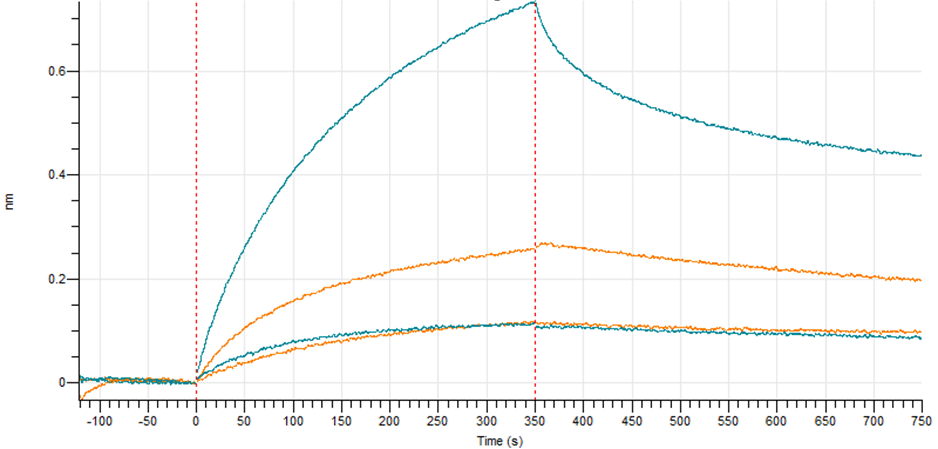
Binding Kinetics and Affinity Characterization
Using the Octet® system, Rockland scientists measure antibody-antigen interactions in real time to determine association rates (ka), dissociation rates (kd), and affinity constants (KD). These measurements provide insight into binding strength and stability, supporting antibody selection and characterization during discovery and assay development.
Figure: Representative BLI sensogram illustrating antibody–antigen binding kinetics measured using the Octet® platform. A series of nine (9) concentrations of mouse anti-antigen antibody (3.0 nM-2.2 µM) were immobilized on an AR2G biosensor and exposed to 20 µg/mL antigen protein. Each colored curve represents the acquired signal of its antibody concentration. The interaction features reproducible duplicate injections and fits to a 1:2 bivalent kinetic model. The values for association constant (ka=32x103M-1s-1) and dissociation constant (kd=8.74x10⁻⁴s-1), and affinity constant (KD=27.3 nM) are depicted in the graph.
Antibody Characterization & Screening Workflows

Epitope Binning Analysis
Bio-Layer Interferometry enables rapid epitope binning to determine whether antibodies bind overlapping or distinct regions of a target antigen. By evaluating competitive binding interactions across antibody panels, researchers can identify groups of antibodies that recognize unique epitopes and differentiate clones with similar binding characteristics.
At Rockland, epitope binning workflows are used to characterize antibody libraries, support therapeutic candidate selection, and identify compatible antibody pairs for diagnostic assay development. The Octet® platform enables high-throughput binning experiments that generate interaction matrices revealing epitope diversity across large antibody panels.
Figure: Representative epitope binning response data showing competitive binding interactions used to classify antibodies into distinct epitope groups. Adapted from the Sartorius Octet® RH96 high-throughput screening application note (access the full study below).
Antibody Pair Screening for Immunoassay Development
BLI can also be used to help rapidly identify antibody binding pairs for use in other assays such as ELISA and lateral flow assays (LFA).
Figure: Screening of candidate antibodies for antigen capture and detection in a sandwich assay. Of the antibodies tested, one antibody demonstrated superior performance compared to the others and was selected for further characterization and development.
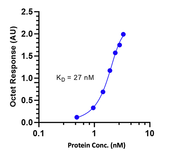
Rapid Protein Quantification Using BLI
Faster and as accurate as ELISA, the Octet® system directly measures the presence of specific proteins and other molecules in solution with minimal interference from complex matrices.
Figure: Steady-state signals (blue-filled symbols) are fitted to a bivalent binding site model (blue line). A 4-parameter non-linear regression curve fit was used for the integration of the data.

Example Application: High-Throughput Screening of H5N1 Hemagglutinin Antibodies
Rockland scientists recently applied Octet® BLI workflows to characterize a large panel of monoclonal antibodies targeting hemagglutinin from highly pathogenic avian influenza H5N1 clade 2.3.4.4b. The study demonstrates how high-throughput BLI can rapidly differentiate antibody binding kinetics and epitope diversity and epitope diversity through high-throughput epitope binning workflows, supporting early candidate selection in discovery programs.
Explore the H5N1 antibody screening applicationWhy Work With Rockland for BLI Analysis
![]()
Antibody Development & Characterization Expertise
Decades of experience in antibody generation, characterization, and assay development enable us to interpret BLI data in the context of real discovery workflows
![]()
High-Throughput Screening Experience
Our scientists routinely apply BLI workflows to rapidly screen antibody libraries, evaluate binding kinetics, and identify differentiated clones
![]()
Actionable Data for Assay Development
From kinetic analysis to antibody pair screening, we generate data that supports assay optimization and downstream research applications
Interested in Bio-Layer Interferometry Services?
Discuss your antibody characterization or assay development needs with our scientists to determine how BLI analysis can support your research program.